
Renal graft parenchyma qualitative assessment through fluorescence angiography before kidney transplantation
Giuseppe Ietto1, Federica Masci1, Davide Brusa1, Giacomo Gaglione1, Domenico Iovino1, Cristiano Parise1, Valentina Iori1, Caterina Franchi1, Andrea Vigezzi1, Fabio Benedetti1, Enrico Ferri1, Andrea Greco1, Martina Pardo1, Matteo Tozzi2, Giulio Carcano1.
1General, Emergency and Transplant Surgery Department, ASST-Settelaghi, Ospedale di Circolo e Fondazione Macchi, Varese, Italy; 2Vascular Surgery Department, ASST-Settelaghi, Ospedale di Circolo e Fondazione Macchi, Varese, Italy
Introduction: Organ shortage is responsible of extensive use of extended criteria donors (ECDs). These grafts require qualitative assessment before transplantation, currently obtained by direct histopathological evaluation through incisional biopsy performed on back-table. Our aim is to develop an alternative way to assess the conditions of renal parenchyma much faster but equally unbiased. To this end, we performed graft Indocyanine green (ICG) angiography with quantitative assessment of fluorescence intensity, during back table surgery.
Materials and Methods: We dissolved 1.66 mg of ICG (Verdye®) in 1L of solution for organ preservation and we performed a fluorescence angiography during renal graft back table. Pictures of the kidneys have been acquired during parenchymal perfusion through a dedicated high definition fluorescent camera (Karl Storz Endoscopes®).
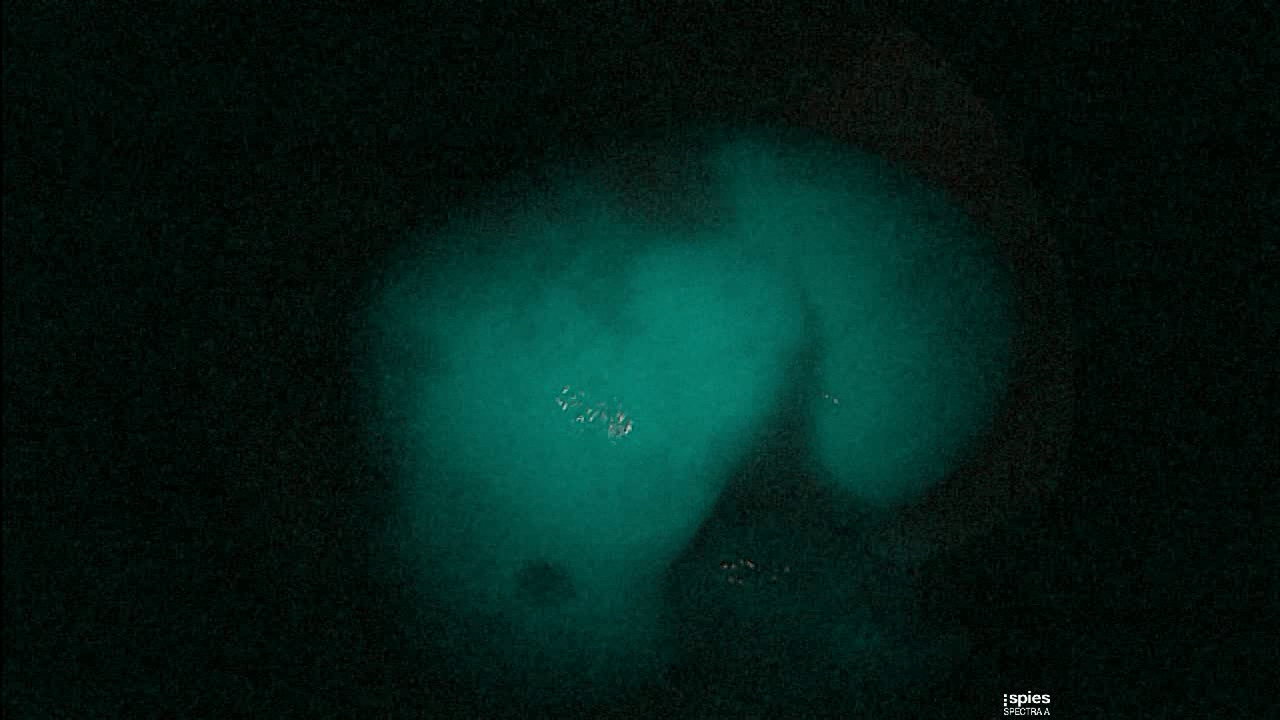
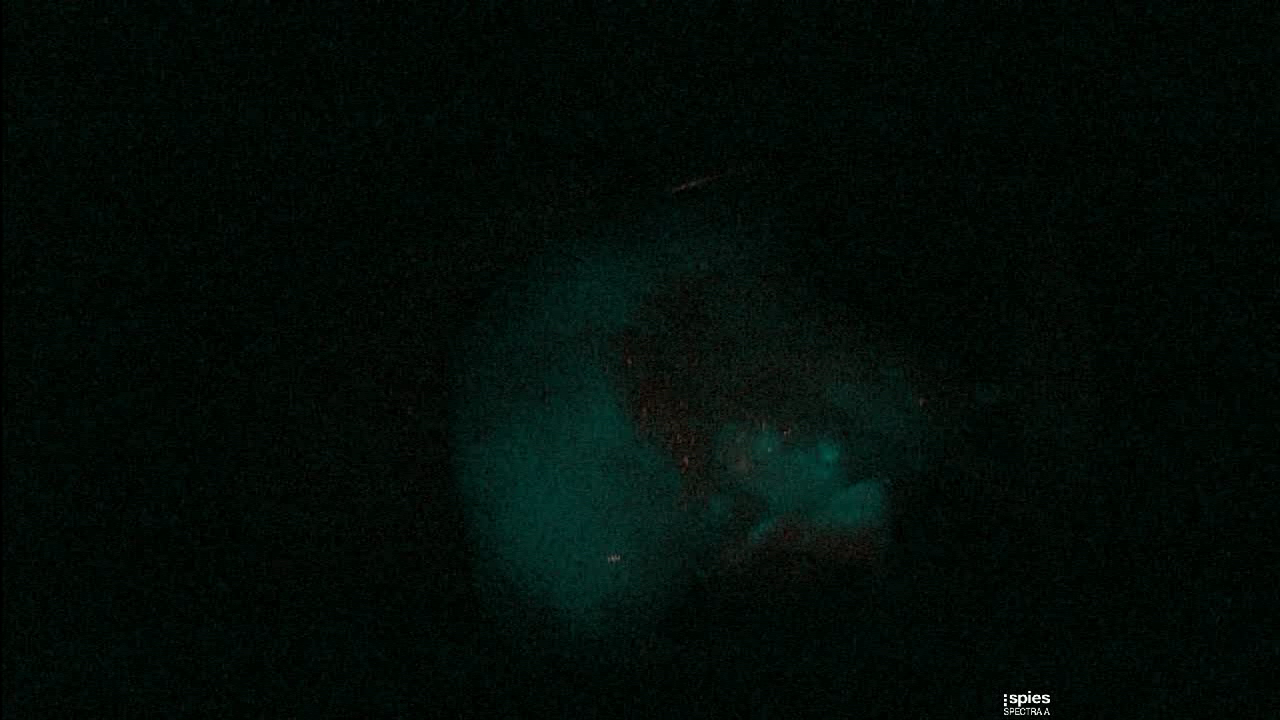
The images were then analyzed using a specific open source software (ICY bioimage analysis®, Quantitative Image Analysis Unit, Institut Pasteur, Paris).

The software calculates the medium intensity of fluorescence in a sample of graft parenchyma on the picture and its homogeneity, and allows defining an Index. This extracted Index was then correlated with transplants outcome, in terms of early recovery vs Delayed Graft Function (DGF).
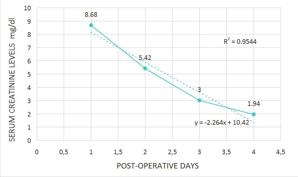
Results and Discussion: The postoperative course of 13 patients were analysed, transplanted in our Transplant Surgery Department between September 2019 and February 2020, with a mean age of 55 years. Of these 4 experienced DGF and 9 experienced an early graft function (EGF). The mean value of the Index for DGF group was 6.75 versus the mean value for EGF group that was 13.17. We did not observed any overlapping between the Index values of the two groups. The Mann-Whitney u-test confirmed a statistically significant difference with a p value < 0.05. We also identified a correlation between our Index and the post-operative decrease of serum creatinine, in terms of reduction trend during the first 4 post-transplant days, with a statistically significant difference.
Considering our Index related with the histopathological analysis in terms of Karpinski score, the results suggest a correlation. In fact, the only graft not suitable for transplant revealed a fluorescence intensity by far lower than the mean value.
Conclusion: Preoperative fluorescent ICG angiography appears safe, easy to use and reproducible. Our first preliminary data suggest a correlation between the Index derived from fluorescence intensity analysis and the outcome of the grafts in terms of early recovery. Larger sample and long term follow-up are obviously required to consider this technique as a valuable option to histopathological evaluation, in order to define the graft suitability for transplant and to predict the postoperative course with the goal of immunosuppressive therapy management.
[1] Friedewald JJ. Utilization and outcomes of marginal kidneys - Using kidney donor risk index to move beyond the current labels. Am J Transplant. 2012;12(8):1971-1972.
[2] Rother U, Gerken ALH, Karampinis I, et al. Dosing of indocyanine green for intraoperative laser fluorescence angiography in kidney transplantation. Microcirculation. 2017;24(8).
[3] Karpinski J, Lajoie G, Cattran D, et al. Outcome of kidney transplantation from high-risk donors is determined by both structure and function. Transplantation. 1999;67(8):1162-1167.
[4] Desmettre T, Devoisselle JM, Mordon S. Fluorescence properties and metabolic features of indocyanine green (ICG) as related to angiography. Surv Ophthalmol. 2000;45(1):15-27.
[5] Rother U, Amann K, Adler W, et al. Quantitative assessment of microperfusion by indocyanine green angiography in kidney transplantation resembles chronic morphological changes in kidney specimens. Microcirculation. 2019;26(3):e12529.
[6] Perico N, Cattaneo D, Sayegh MH, Remuzzi G. Delayed graft function in kidney transplantation. Lancet. 2004;364(9447):1814-1827.
There are no comments yet...